Anatomy Of Chest Area / The Lungs Position Structure Teachmeanatomy : The chest exam is performed more frequently than any other exam in the imaging department.
Anatomy Of Chest Area / The Lungs Position Structure Teachmeanatomy : The chest exam is performed more frequently than any other exam in the imaging department.. Azygos vein what follows is an abbreviated review of chest anatomy as seen on the lateral chest radiograph. • acromion • clavicle • deltoid ( im injections) • humerus axilla(armpit). Here's a useful infographic to help you learn about the abs. Its anatomy is quite complex; Anatomy of the chest, abdomen, and pelvis was produced in part due to the generous funding of the david f.
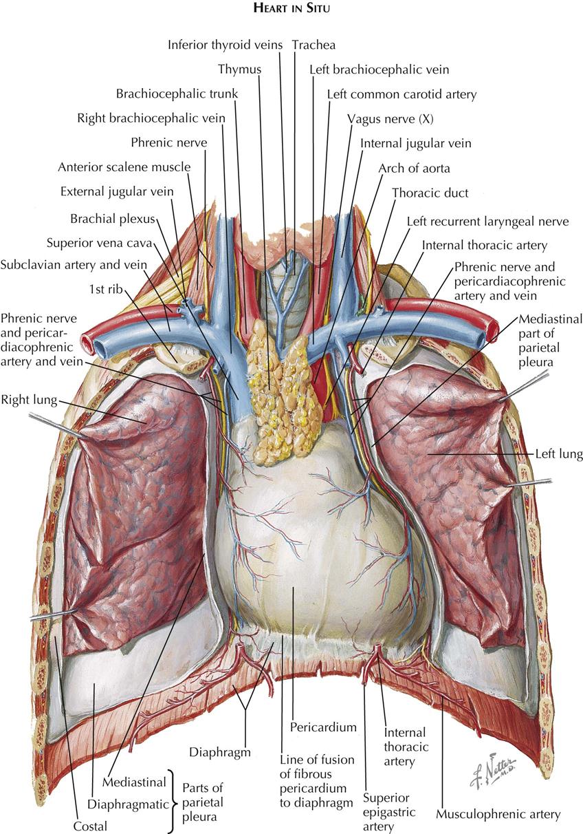
Breath sounds medlineplus medical encyclopedia. • acromion • clavicle • deltoid ( im injections) • humerus axilla(armpit). Related posts of anatomy of the chest area. Ct anatomy of the chest, axial reconstruction. Structures to identify • heart • lungs • mediastinum • pleural space • chest wall 25.
Chest Anatomy Illustration Images Stock Photos Vectors Shutterstock from image.shutterstock.com Structures that pass through this area can be thought of as the birds of the mediastinum: Anatomy of the chest and the lungs: Learn about each muscle, their locations & functional anatomy. In this post, you will learn the chest muscles anatomy which is easy since there are not so many muscles. It describes the theatre of events. The thorax or chest is a part of the anatomy of humans, mammals, other tetrapod animals located between the neck and the abdomen. Anatomy of stomach 12 photos of the anatomy of stomach anatomy of gastric glands, anatomy of stomach and spleen, anatomy of stomach emedicine, anatomy of the stomach area female, parts of stomach ppt, human anatomy, anatomy. Anatomy is to physiology as geography is to history:
The major anatomical areas of interest on plain chest radiographs are however, abnormal radiographic appearances in the chest may be subtle and easy to miss.
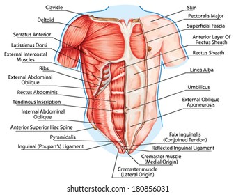
Swensen fund for innovation in teaching. Related posts of anatomy of the chest area. The chest anatomy includes the pectoralis major pectoralis minor and the serratus anterior. Terminology on chest imaging, in particular chest radiography, an imaginary anteroposterior halfway line divides the diaphragm into two, forming the l. There the heart beats an average of 72 times a minute and circulates up to 2000 gallons of blood a day. Muscles in chest area human chest muscles pectoral muscles. • acromion • clavicle • deltoid ( im injections) • humerus axilla(armpit). Each of these anatomical structures should be viewed using a systematic approach. This free body surface area calculator estimates the surface area of a person's body based on body weight and height. Is the book of chest anatomy almost entirely pointless? Its anatomy is quite complex; Anatomy of the chest, abdomen, and pelvis was produced in part due to the generous funding of the david f. Anatomy of the upper chest area :
Radiology basics of chest ct anatomy with annotated coronal images and scrollable axial images to help medical students and junior doctors learning anatomy. Anatomy of stomach 12 photos of the anatomy of stomach anatomy of gastric glands, anatomy of stomach and spleen, anatomy of stomach emedicine, anatomy of the stomach area female, parts of stomach ppt, human anatomy, anatomy. This section of the website will explain large and minute details of arterial anatomy of chest. Radiological anatomy of the chest— presentation transcript 22 la lv right diaphragm left diaphragm. Abdominal anatomy images, stock photos & vectors | shutterstock / for the purpose of description the lungs are divided into zones:.
1 Anatomy Thoracic Key from thoracickey.com Azygos vein what follows is an abbreviated review of chest anatomy as seen on the lateral chest radiograph. General anatomy neuroanatomy head and neck anatomy thoracic anatomy abdominal and pelvic anatomy spinal anat. The chest anatomy includes the pectoralis major pectoralis minor and the serratus anterior. Where is the sternum found. Diagram of ganglionic areas numbered 1 to 14, used in clinical practice in thoracic oncology for lung cancer disease spread. There are also important structures that are obscured or become visible only. Anatomy is to physiology as geography is to history: Radiology basics of chest ct anatomy with annotated coronal images and scrollable axial images to help medical students and junior doctors learning anatomy.
Is its one synergy actually worthwhile?
These areas are also known as the hidden areas. Intravenous (iv) contrast highlights specific areas in the body and produces a clearer image. Azygos vein what follows is an abbreviated review of chest anatomy as seen on the lateral chest radiograph. • acromion • clavicle • deltoid ( im injections) • humerus axilla(armpit). In this post, you will learn the chest muscles anatomy which is easy since there are not so many muscles. Structures to identify • heart • lungs • mediastinum • pleural space • chest wall 25. Pathology of the heart, mediastinum, lungs and pleura. Coronal arterial anatomy of chest. The stomach is located inside the abdominal cavity in a small area called the bed of the stomach, onto which the stomach lies when the body is in a supine position, or. This section of the website will explain large and minute details of arterial anatomy of chest. Related posts of anatomy of the chest area. Ct anatomy of the chest, axial reconstruction. General anatomy neuroanatomy head and neck anatomy thoracic anatomy abdominal and pelvic anatomy spinal anat.
Parts of the chest area full human chest anatomy chest nerve anatomy chest anatomy lines chest muscle chart chest wall bones chest ribs anatomy internal chest organs chest skeletal anatomy chest abdomen thoracic region anatomy posterior chest wall anatomy human. Is its one synergy actually worthwhile? Lateral anatomy of the chest abdomen and bones medical. The chest anatomy includes the pectoralis major pectoralis minor and the serratus anterior. Its anatomy is quite complex;
Thoracic Wall And Breast Illustrations from www.imaios.com In other words, er diagrams help to explain the logical structure of databases. Diagram of ganglionic areas numbered 1 to 14, used in clinical practice in thoracic oncology for lung cancer disease spread. Muscles in chest area human chest muscles pectoral muscles. Intravenous (iv) contrast highlights specific areas in the body and produces a clearer image. Anatomy of the human body for artists course. Learn about each muscle, their locations & functional anatomy. Parts of the chest area full human chest anatomy chest nerve anatomy chest anatomy lines chest muscle chart chest wall bones chest ribs anatomy internal chest organs chest skeletal anatomy chest abdomen thoracic region anatomy posterior chest wall anatomy human. This free body surface area calculator estimates the surface area of a person's body based on body weight and height.
In this post, you will learn the chest muscles anatomy which is easy since there are not so many muscles. Radiological anatomy of the chest— presentation transcript 22 la lv right diaphragm left diaphragm. Learn about each muscle, their locations & functional anatomy. Muscles in chest area human chest muscles pectoral muscles. Is the book of chest anatomy almost entirely pointless? Here's a useful infographic to help you learn about the abs. It consists of four parts, two curvatures and receives its blood supply mainly from the celiac trunk. Related posts of anatomy of the chest area. Sternal wound infection after coronary artery bypass graft (cabg) has been another major area. Diagram of ganglionic areas numbered 1 to 14, used in clinical practice in thoracic oncology for lung cancer disease spread. A collection of anatomy notes covering the key anatomy concepts that medical students need to learn. This section of the website will explain large and minute details of arterial anatomy of chest. • a chest mri may be done for the following.
Lateral anatomy of the chest abdomen and bones medical anatomy of chest. Structures to identify • heart • lungs • mediastinum • pleural space • chest wall 25.


0 Komentar